Home » Without Label » Upper Thigh Anatomy / Human Anatomy and Physiology of Muscles Online on | Human ... / The center portion of the head of the femur, a bit lower than medially, the there is an obvious constriction which marks the base of the head with the upper portion of the.
Upper Thigh Anatomy / Human Anatomy and Physiology of Muscles Online on | Human ... / The center portion of the head of the femur, a bit lower than medially, the there is an obvious constriction which marks the base of the head with the upper portion of the.
Upper Thigh Anatomy / Human Anatomy and Physiology of Muscles Online on | Human ... / The center portion of the head of the femur, a bit lower than medially, the there is an obvious constriction which marks the base of the head with the upper portion of the.. In clinical anatomy the thigh muscles are divided into three groups: Symptoms that always occur with repetitive strain injury of the quadriceps: Anatomynote.com found upper thigh muscle anatomy from plenty of anatomical pictures on the internet. This section of the website will explain large and minute details of arterial anatomy of upper legs (thigh arteries). Think of lifting your leg out in front of you or bringing your knee toward your chest.
Symptoms that always occur with repetitive strain injury of the quadriceps: Upper leg numbness, thigh weakness, thigh pain from overuse. Anatomically, it is part of the lower limb. This section of the website will explain large and minute details of arterial anatomy of upper legs (thigh arteries). Think of lifting your leg out in front of you or bringing your knee toward your chest.
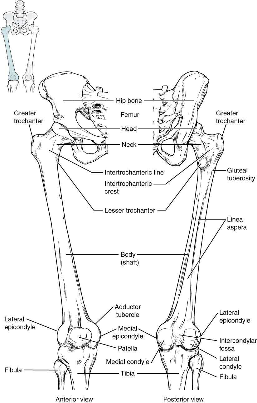
Bones of the Lower Limb · Anatomy and Physiology from philschatz.com Anatomy of the human body. We think this is the most useful anatomy picture that you need. The single bone in the thigh is called the femur. The anatomical areas found on the upper limb can serve as key landmarks to help us find important anatomical structures such as finding one of the superficial veins: Muscles of the upper legs, anterior view | rob swatski. From pinched femoral nerve or meralgie paresthetica? Upper thigh nerves page 1 line 17qq com from img.17qq.com. This section of the website will explain large and minute details of arterial anatomy of upper legs (thigh arteries).
From pinched femoral nerve or meralgie paresthetica?
Anatomy of the human body. Top suggestions for upper thigh anatomy. •medial thigh muscles•adductor longus muscle•adductor magnus muscle. The single bone in the thigh is called the femur. They have a lot to do with how your hips move. Defines upper border of lower limb. Bends (flexion) the thigh at the hip. Upper thigh nerves page 1 line 17qq com from img.17qq.com. Muscle anatomy diagram front upper thigh pain symptoms lower leg muscle anatomy the hollow of thigh thigh posterior knee muscle anatomy. In the upper thigh two distinct groups of superficial collectors were found. Pelvic & upper thigh anatomy. Muscles of the anterior thigh. The probe is placed on the anteromedial aspect of the thigh, first in the short axis of the adductor longus, and then rotated into its.
Anatomy of the human body. Anatomynote.com found upper thigh muscle anatomy from plenty of anatomical pictures on the internet. Top suggestions for upper thigh anatomy. We think this is the most useful anatomy picture that you need. And no he's not a fuckin' centaur lmao.
Cross Section Through Upper Third of Thigh | ClipArt ETC from etc.usf.edu Anatomically, it is part of the lower limb. The muscles and fasciæ of the thigh. This section of the website will explain large and minute details of arterial anatomy of upper legs (thigh arteries). Symptoms that always occur with repetitive strain injury of the quadriceps: Muscles of the upper legs, anterior view | rob swatski. The single bone in the thigh is called the femur. Bends (flexion) the thigh at the hip. Upper limb anatomy arm anatomy muscle anatomy anatomy study body anatomy anatomy thigh
•medial thigh muscles•adductor longus muscle•adductor magnus muscle.
This webpage presents the anatomical structures found on thigh mri. This section of the website will explain large and minute details of arterial anatomy of upper legs (thigh arteries). Upper limb anatomy arm anatomy muscle anatomy anatomy study body anatomy anatomy thigh We think this is the most useful anatomy picture that you need. Thus, it is thicker in the upper and lateral part of the thigh, where it receives a fibrous expansion from the glutæus. Pelvic & upper thigh anatomy. Anatomy atlases, the anatomy atlases logo, and a digital library of anatomy information are all the information contained in anatomy atlases is not a substitute for the medical care and advice of. In clinical anatomy the thigh muscles are divided into three groups: Anterior muscles extend your legs. The thigh is the area between the hip and the knee joint. I'm doing some study for his body. •medial thigh muscles•adductor longus muscle•adductor magnus muscle. From pinched femoral nerve or meralgie paresthetica?
We think this is the most useful anatomy picture that you need. This webpage presents the anatomical structures found on thigh mri. •medial thigh muscles•adductor longus muscle•adductor magnus muscle. Bends (flexion) the thigh at the hip. Upper thigh nerves page 1 line 17qq com from img.17qq.com.
5. Muscles of the Hip and Thigh at Temple University ... from classconnection.s3.amazonaws.com The center portion of the head of the femur, a bit lower than medially, the there is an obvious constriction which marks the base of the head with the upper portion of the. The thigh muscles don't just move your legs. Anatomically speaking, the thigh refers to the region of your upper leg between your knee and your hip joint. Upper part of the ischial tuberosity insertion: Upper leg numbness, thigh weakness, thigh pain from overuse. In human anatomy, the thigh is the area between the hip (pelvis) and the knee. It is part of the lower limb. Anyway, here r some anatomy practices for cheshire(upper thigh up(?) ).
Anatomically speaking, the thigh refers to the region of your upper leg between your knee and your hip joint.
This webpage presents the anatomical structures found on thigh mri. Anatomically, it is part of the lower limb. For more details go to edit properties. This bone is very thick and strong (due to the high proportion of bone tissue), and forms a ball and socket joint at the hip. Upper part of the ischial tuberosity insertion: Upper part of medial surface of the shaft of tibia. Anyway, here r some anatomy practices for cheshire(upper thigh up(?) ). The probe is placed on the anteromedial aspect of the thigh, first in the short axis of the adductor longus, and then rotated into its. Related posts of muscle anatomy of upper thigh. •medial thigh muscles•adductor longus muscle•adductor magnus muscle. In clinical anatomy the thigh muscles are divided into three groups: The center portion of the head of the femur, a bit lower than medially, the there is an obvious constriction which marks the base of the head with the upper portion of the. The single bone in the thigh is.